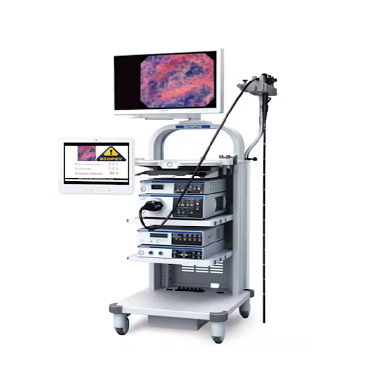
CV-1500
- Point of origin
-
FOB Hongkong
- Testing item
-
Gastrointestinal
- Application scenarios
-
Hospital
Unified Platform with 5 LED Spectrum Technology
Broad Compatibility
Enhanced Observations
The CV-1500 provides several advanced imaging modes in addition to standard white light and conventional NBI (Narrow Band Imaging) and AFI (Auto Fluorescence Imaging):
TXI (Texture & Color Enhancement Imaging): Enhances the mucosal surface by optimizing its structure, color tone, and brightness, facilitating more precise diagnostics.
RDI (Red Dichromatic Imaging): Enhances visibility of deep blood vessels and bleeding points, aiding in the detection of underlying issues.
BAI-MAC (Brightness Adjustment Imaging with Maintenance of Contrast): Improves brightness in darker areas while maintaining contrast, ensuring clearer visuals in challenging conditions.
Intuitive, User-Friendly Functions
The CV-1500 features a One-Touch Connector for swift and simple setup, eliminating the need for white balance adjustments. This streamlines the workflow and reduces procedure times. The touch-sensitive panel allows for intuitive operation, and convenient features like Pre-freeze and MyCV mode enhance user experience. Additionally, the LED bulbs used in the system have a long lifespan, reducing the need for frequent replacements and minimizing downtime.
The clinical application of the CV-1500 Video System 4K HD—Gastrointestinal Endoscopy System in disease diagnosis is significant due to its advanced imaging capabilities.
Clinical Applications:
Detection and Characterization of Lesions:
White Light Imaging (WLI): Provides high-resolution images to detect surface lesions, polyps, and abnormalities.
Narrow Band Imaging (NBI): Enhances visualization of mucosal structures and capillary networks, aiding in the identification of neoplastic changes.
Evaluation of Inflammatory Diseases:
Assessment of Vascular Abnormalities:
Red Dichromatic Imaging (RDI): Improves visibility of deep blood vessels and bleeding points, crucial for diagnosing conditions like gastrointestinal bleeding, angiodysplasia, and varices.
Cancer Detection and Screening:
Auto Fluorescence Imaging (AFI): Differentiates between normal and abnormal tissue based on fluorescence, enhancing early detection of gastrointestinal cancers, including esophageal, gastric, and colorectal cancers.
TXI and NBI: Provide detailed visualization that aids in identifying dysplasia and early-stage malignancies.
Guidance for Therapeutic Procedures:
Brightness Adjustment Imaging with Maintenance of Contrast (BAI-MAC): Enhances brightness in darker portions of the image while maintaining contrast, improving the accuracy of therapeutic procedures such as polypectomy, endoscopic mucosal resection (EMR), and endoscopic submucosal dissection (ESD).
Improved Workflow and Efficiency:
Specific Disease Applications:
Gastroesophageal Reflux Disease (GERD): Enhanced imaging helps in the evaluation of esophagitis and Barrett’s esophagus.
Peptic Ulcer Disease: Improved visibility of ulcers and surrounding mucosa.
Colorectal Cancer: Early detection of polyps and malignancies through enhanced imaging techniques.
Inflammatory Bowel Disease (IBD): Detailed assessment of mucosal inflammation and ulceration.
Gastrointestinal Bleeding: Accurate localization of bleeding sources.
| Rated voltage | 100-240 V AC; Within 土10% |
| Frequency | 50/60 Hz; within +3 Hz |
| Rated input | 600 VA |
| Dimensions (WxHx D) | 370x 198 x 488 mm; 398 x 218 x 580 mm (maximum) |
| Weight | 19.4 kg |
| Type of protection against electric shock | Class |
| Degree of protection against electric | Depend on applied part. (The degree of protection against electric shock of this product is BF type if the mounting part to be |
| shock of applied part | connected to this product is BF type. However CF type is not subject to combination in this product,) |
| Degree or protection against explosion | The video system center should be kept away from flammable gases. |
| Analog signal output | VBS composite |
| Digital signal output | 12G-SDI (SMPTE ST 2082), 3G-SDI (SMPTE424M), HD-SDI (SMPTE292M), SD-SDI (SMPTE259M) |
| User settings | The function settings for up to 20 users can be stored. |
| Color tone adjustment | Adjust the color tone of each endoscopic image for White light observation mode, NBI observation mode, and RDI observation mode. . Red adjustment: +8 steps : Blue adjustment:土8 steps . Chroma adjustment: +8 steps |
| Automatic gain control (AGC) | The image can be electronically amplified when the light is inadequate due to the distal end of the endoscope being too far from the object. |
| Contrast | . H(High) : Darkens the dark part and brightens the bright part. . L (Low): Brightens the dark part and darkens the bright part. |
| BAI-MAC | Brightness adjustment with maintenance of contrast |
| Iris | The iris modes can be switched. . Auto: The brightness is adjusted based on the brightest part of the central part and the average brightness of the periphery part. . Peak : The brightness is adjusted based on the brightest part of the endoscopic image. . Average: The brightness is adjusted based on the average brightness of the endoscopic image.Fine patterns or edges in the endoscopic images can be enhanced electrically to increase the image sharpness. |
| Switching the enhancement modes | The enhancement level can be selected from 3 levels (OFF, 1, 2, and 3) |
| Image size selection | The size of the endoscopic image can be selected from 2 modes. (Except SDTV) |
| Electric zoom | Switch between mode 1, mode 2, and mode 3. |
| PIP/POP | Switch between PIP and POP. |
| Aspect ratio | Switch between 16:9 and 4:3. (Except SDTV) |
| Freeze Pre-freeze | Freeze the endoscopic image. The image with the least blur is selected from the images captured in the set time period before freeze operation and displayed. |