At the end of 2016, scientists analyzed pre-pregnancy and postpartum imaging data and discovered for the first time that pregnancy can cause significant changes in brain structure, especially a notable reduction in the volume of gray matter areas related to cognition.
However, these studies only helped scientists understand the differences in brain structure before and after pregnancy. How the brain structure changes throughout the pregnancy process remained unknown.
Recently, a research team led by Laura Pritschet and Emily G. Jacobs from the University of California, Santa Barbara, and Elizabeth R. Chrastil from the University of California, Irvine, published a significant study in the renowned journal Nature Neuroscience, closing this gap in understanding.
They conducted an extensive pregnancy imaging study on a healthy first-time mother and found that, as gestation progresses, the gray matter volume of the cerebral cortex decreases, cortical thickness reduces, and the volume of multiple hippocampal regions also declines. However, the integrity of white matter microstructure improves.
Notably, the reduction in gray matter volume and cortical thickness continues for two years postpartum, while the improvement in white matter microstructure returns to baseline levels soon after delivery—changes that had been difficult to detect in previous studies.
This is the first time scientists have revealed how the female brain changes throughout pregnancy.
The study was made possible by a 38-year-old mother who began undergoing MRI scans three weeks before her pregnancy. Her pregnancy lasted 36 weeks, during which she underwent 19 MRI scans. She was followed for two years postpartum, during which she had an additional 7 MRI scans. Throughout the study, nearly 20 venous blood samples were also collected.
Blood test results showed that serum hormone (17β-estradiol and progesterone) concentrations increased significantly during pregnancy and dropped sharply after delivery.
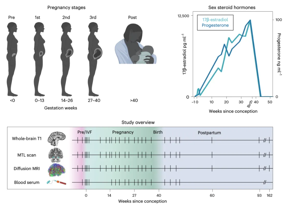
Overall, cortical gray matter volume (GMV), cortical thickness (CT), and total brain volume decreased linearly throughout pregnancy, with a partial rebound after delivery. As mentioned earlier, the cerebral cortex gray matter is closely related to social cognition and thought processes, yet during pregnancy, gray matter volume decreased.
In contrast, the microstructural integrity of white matter increased during the first six months of pregnancy and returned to baseline levels after delivery. White matter, located deep within the brain, facilitates communication between different brain regions.
Additionally, the lateral ventricles and cerebrospinal fluid expanded in a nonlinear pattern during the second half of pregnancy, then sharply decreased after delivery.
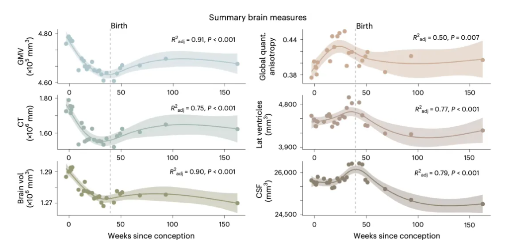
Pritschet's team focused on the 19 MRI scans taken before and during pregnancy, finding a positive correlation between total brain volume, gray matter volume (GMV), and cortical thickness (CT), while the lateral ventricles and cerebrospinal fluid were negatively correlated with gray matter volume (GMV).
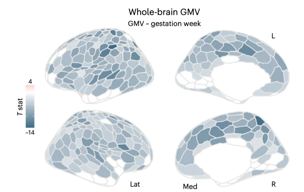
They also noticed that changes in gray matter volume (GMV) were nearly universal throughout the cerebral cortex, with 80% of the 400 brain regions of interest showing negative correlations with gestational age, indicating that the volume of the cerebral cortex decreases almost continuously throughout pregnancy.
Remarkably, the reduction in gray matter volume (GMV) was significantly correlated with serum estradiol and progesterone concentrations, and similar findings were observed in cortical thickness (CT) changes related to pregnancy.
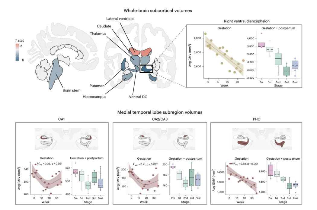
In addition to the widespread reduction in cortical gray matter volume (GMV), several subcortical regions also experienced significant volume decreases during pregnancy, including the bilateral ventral diencephalon (e.g., hypothalamus, substantia nigra, mammillary bodies, lateral geniculate nucleus, and red nucleus), caudate nucleus, hippocampus, thalamus, and brainstem.
Specifically, in the hippocampus, the volume of CA1, CA2, and CA3 regions decreased nonlinearly during pregnancy, while the volume of the parahippocampal cortex (PHC) decreased linearly.
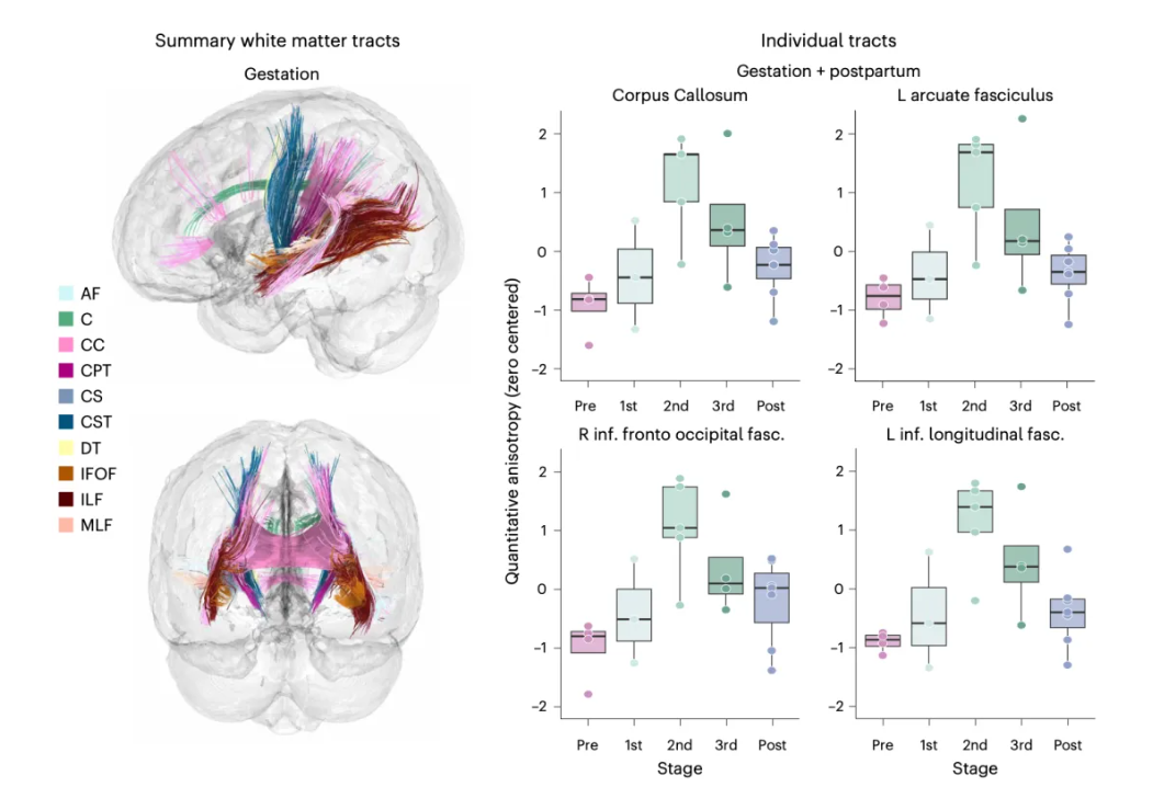
Beyond the brain shrinkage, Pritschet's team observed that the microstructural integrity of white matter fiber tracts increased during pregnancy, which was synchronized with rising 17β-estradiol and progesterone levels. This change in white matter suggests increased communication between brain regions, potentially related to maternal behavior.
At the end of the study, Pritschet's team compared the participant's data with brain imaging data from eight other subjects. They found that the changes in gray matter volume (GMV) throughout pregnancy were far greater than normal brain variability. On average, cortical gray matter volume (GMV) changes were nearly three times higher than in the control group scanned over a similar period.Overall, the research led by Laura Pritschet, Emily G. Jacobs, and Elizabeth R. Chrastil fills a research gap on how pregnancy affects the female brain, advancing our understanding of human brain plasticity. However, this study was based on a single participant, and further human data are needed to confirm these findings.
Additionally, this study only documented brain changes during pregnancy and could not explain how these changes relate to behavior. Future research may need to investigate this question using animal models.